Dental radiography is necessary to help your dentist visualize and assess your teeth, bone, jaw joints, nerves, sinuses, and other orofacial landmarks. In addition to high-quality 2-dimensional dental X-rays, the team at Well Rooted Dentistry also utilizes cone beam computed tomography (CBCT), which provides a comprehensive, 3-D view of your oral anatomy. Here, we’ll explain how this technology works, when it is recommended, and the benefits it offers.
What is cone beam computed tomography?
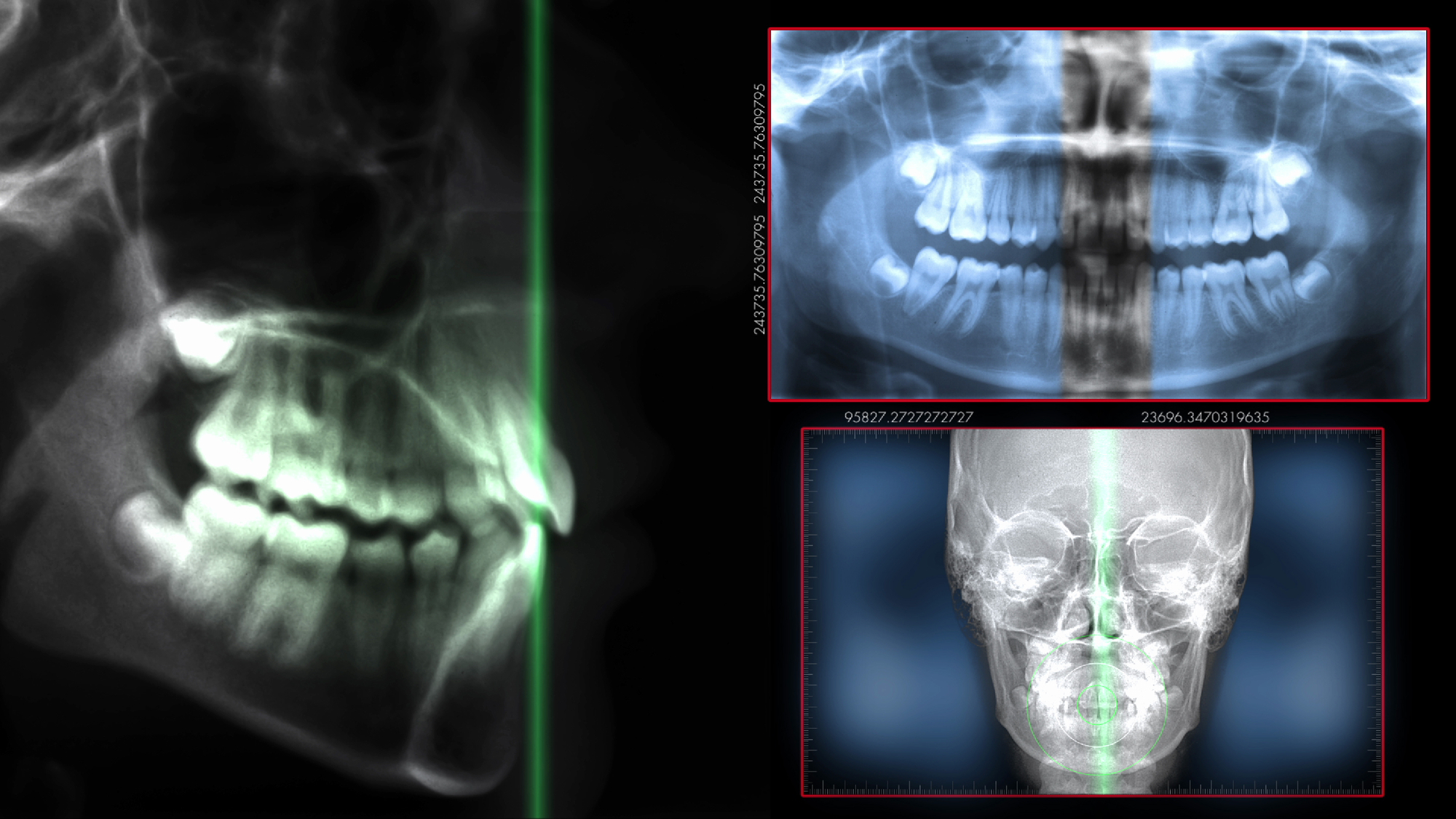
Unlike a 2-D periapical or bitewing X-ray, which only captures one image, a dental CT scan takes hundreds of images, then stitches them together during a single scan. In addition to providing optimal visibility, this type of scan allows your dentist to manipulate the images for an even clearer view. CBCT scans have revolutionized the way that we do dentistry. This technology allows your dentist to obtain more accurate records, and in turn, provide you with a more customized treatment plan.
How is a cone beam scan performed?
CBCT is similar to a traditional CT scan. However, because it focuses on the oral cavity, you do not have to lie in a conventional CT machine. Instead, the patient sits or stands while an X-ray tube rotates around the head. You must remain still while the scan is in progress. The entire process takes about a minute or less to complete. The images are then uploaded directly to our computer software program.
When is cone beam computed tomography recommended?
Cone beam technology has a wide range of uses in dentistry, from cosmetic and restorative treatments to orthodontics and prosthodontics. Your dentist may recommend a cone beam scan when diagnosing and treating a number of conditions, including:
- Missing teeth
- Bone loss
- Orthodontic misalignment
- Decayed or damaged teeth
- TMJ pain
Cone Beam Technology and Dental Implants
CBCT is incredibly useful for dental implant treatment planning. Once the images have been captured, your dentist can determine which location will be optimal for your implant placement. Cone beam scans tell us how much room is available, whether the jawbone is healthy, and the exact locations of your mandibular nerve and maxillary sinuses. Your dentist can even superimpose virtual implants onto the cone beam image. This helps your team know what size and type of dental implant you will need before your surgery even begins.
Prior to the advent of CBCT, dentists had to rely solely on 2-D X-rays. Today, 3-D cone beam technology effectively takes the guesswork out of treatment planning.
What are the advantages of cone beam computed tomography in dentistry?
Cone beam technology offers a number of benefits in dentistry. This type of scan offers:
- Enhanced accuracy and image quality: In addition to optimal accuracy, CBCT scans offer the highest-quality images available.
- Views of both hard and soft tissues: While conventional X-rays only capture bone, CBCT captures both bone and oral soft tissues, allowing for more precise treatment.
- Minimal radiation exposure: CBCT emits less radiation than traditional CT scans, keeping patients safe from overexposure.
- Customizable scans: Dental CT scans can capture the entire dentition, or just a portion of the jaw, depending on the patient’s unique circumstance. In other words, we can gain better insights into your oral health without requiring multiple scans.
Learn more about our advanced technology.
To learn more about cone beam technology, or any of the services we provide, contact our Providence, RI practice online anytime. You can also call our office at (401) 533-9680.